- Cerebral Malaria – Symptoms - March 16, 2020
- Rabies a deadly disease: a must read article - January 6, 2020
- Pathological Changes in Malaria - December 30, 2019
Last Updated on January 14, 2024 by The Health Master
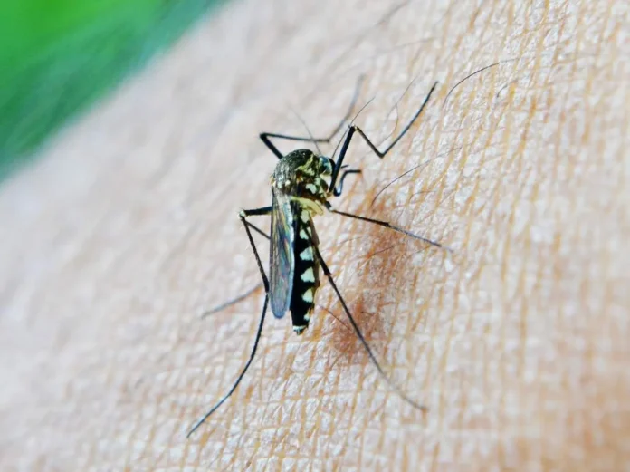
We discuss here about Pathological Changes in Malaria. We have already discussed the cause of Malaria by the bite of infected female Anopheles mosquito. Rarely it can be caused by the infected blood transfusion.
The species which cause malaria are
- P. Vivex
- P.Ovale
- P. Malarae
- P. Falciparum
Pathological Changes in Malaria
- Liver:
- Grossly enlarged. Slate grey or blackish due to deposition of malarial pigments
Microscopically:
- Central veins and Sinusoids are congested in liver.
- Kuffer cells are ladden with blackish haemozoin pigments
- Spleen:
- After a chronic course of malaria the viscearas such as spleen which is partially producing abc and immunity cells, becomes fibrotic and bristle. The capsule becomes thick. The spleen shows blackish pigments within the phagocytes.
- The name malaria was given around 1753 (AD) and its treatment was discovered in the middle of the 17th century.
- Haemozoin pigment ( the malarial pigment) microscopically looks like foramline and melanin pigment
To differentiate the foramline and malarial pigments can be bleached by alchoholic picric acid but the melanin does not get bleached by alchoholic picric acid-melanin is the pigment found in skin in blackish people.
There is nil or less amount in white skinned people.
Other symptoms and more will be discussed in another topic.
Also read other articles of the Author:
1. Malaria – Aeitiology & Symptoms
2. Bombay Blood Group


